

Using Diagnostic Ultrasound (MSKUS) To Take Your Clinic To Next Level Care With Bart McDonald, PT, DPT
Diagnostic ultrasound or musculoskeletal ultrasound (MSKUS) has been used for decades in the healthcare industry. It is also within the scope of practice for almost all PTs in the U.S. However, very few PTs have taken the effort to learn how to use it because it is difficult to learn. But its capabilities, if utilized, are able to significantly improve the care and results that PTs get with their patients. And if PTs truly see themselves as potential gatekeepers for musculoskeletal conditions they MUST have such objective tests at their disposal. It’s the only way to grow in your profession. Bart McDonald , PT has implemented MSKUS in his PT practice to great success, setting his diagnostic ability and patient care head and shoulders above other clinics. Join your host Nathan Shields as he talks to Bart McDonald about how he’s using MSKUS successfully. Find a mentor and start practicing MSKUS today!
—
Listen to the podcast here
Using Diagnostic Ultrasound (MSKUS) To Take Your Clinic To Next Level Care With Bart McDonald, PT, DPT
I’ve got a returning guest, Bart McDonald, the crazy guy from Idaho. Bart, thanks for joining me again.
Nathan, it is so fun to be a part of this. This is a cool thing for physical therapists to be able to glean from each other what’s going on and how to improve the practice of Physical Therapy.
That’s the whole reason why I’m doing this. I love having successful PT owners like you on. For those who didn’t capture my episode with Bart McDonald , he is the CEO and Founder of Superior Physical Therapy and Diagnostics in Idaho. He has multiple clinics. In the previous episode that we did, we talked about the book that he had written called Debt-Free PT and how to help PTs get out from underneath the burden of student debt.
Also, how owners can help those new grads who are underneath a heavy student debt load, you can go back to that and glean some information on how to help our new grads get out from under the student loan burden that they have and how we can incentivize them, talk to them, and recruit them as well.
We’re talking about something different because those who have followed this show know that I have done diagnostics and I do diagnostics. I’m board-certified in electromyography. I have my ECS. I have also learned how to work on musculoskeletal ultrasound, although I’m not fellowship-trained and I don’t have my certificate in it. Nonetheless, I know plenty of owners that do across the country. Bart is one of the owners that I know through our hands-on diagnostics network that has been successful with the implementation of musculoskeletal ultrasound in his practices.
I wanted to highlight him and talk about musculoskeletal ultrasound because he feels like I do that musculoskeletal ultrasound should be in the majority of outpatient orthopedic clinics across the country. It should be a widely used tool for the benefit of physical therapists in general and a benefit and an addition to the care that we can provide based on the diagnostics that we can do within our scope of practice. Correct me if I’m wrong, Bart. Most states allow for physical therapists to do a diagnostic ultrasound.
That’s my understanding. When we first got into it, I looked at our practice, and I think that’s where everybody should go. I was looking at EMG. In the state of Idaho, we have a great solid Practice Act for us being able to do nerve conduction studies and EMG. We looked at ultrasound and ultrasound was all over our Practice Act.
You talked about using acoustic energy both for the treatment and/or benefit of the patient. Diagnostical sounds have been around since the ‘50s. As technology has improved, it’s much better. Who knows exactly what those folks we were all thinking about when they were talking about acoustic energy and put the Idaho Practice Act together, but it flows well.
From my perspective, I think about doing a PT evaluation. I walked in with a patient and they’ve got shoulder pain. The evaluation from the doctor says, “Shoulder pain.” I perform a number of special tests and I check the range of motion. I’m going to see if they’ve got impingement. I’m going to see if they’ve got any instability or laxity. I’m screening the labrum. I’m checking all of these things, and then I look at the sensitivity and specificity of those special tests I’m using and it’s low. It’s not great.
I remember being in physical therapy school and going, “The sensitivity and specificity are terrible for a lot of our special tests. What should I do with this?” Maybe if I do 50,000 special tests for each shoulder patient, I can get this clinical picture that helps me. That’s what I’ve always done. I can tell you how we started in musculoskeletal ultrasound.
I bring that up because when we compare the sensitivity and specificity of musculoskeletal ultrasound as compared to an MRI, it’s pretty strong. MRI is the gold standard and it varies by joint. I can’t get deep to the ACL well with musculoskeletal ultrasound at the knee, but I can see a lot of structures, the meniscus and all these. Certainly, they have great views of the rotator cuff on the shoulder, all the ligaments of the ankle, etc.

When we talk about the utilization of musculoskeletal ultrasound in a PT practice, I think of it as that extra extension of what I’m using in my clinical exam. It’s another special test for me. Only, it’s a special diagnostic test with some great correlation and great reliability. I can do it time and time again. I can show the patient even in a sequential way that by repeating this every 4 to 6 weeks, depending on the problem, we’re improving beyond, “Did their strength improve? Can they sleep better at night? Did this special test improve slightly? Did their manual muscle testing move up a half grade?” We can look at the tissues and get to the base of their pain and see improvement. It’s phenomenal.
I’ve been saying this for years and it hasn’t come to fruition yet but in the next decades, ultrasound should be like dry needling is now. I’m hoping that it is. You and I have been practicing for probably about the same amount of years but 10 to 15 years ago, we weren’t quite sure what dry needling was. It was way out there.
Ultrasound should even be a greater tool diagnostically. It could have a greater impact on our plans of care than dry needling. It’s something that a lot of physical therapists should embrace to the point where I wish the APTA would do more in supporting diagnostic ultrasound and pushing it more. I don’t know about you, but APTA magazine had a long article about states across the country that we had had grassroots efforts to allow physical therapists to refer for X-rays. There’s no mention in there whatsoever that we can do the diagnostic ultrasound.
I sent a letter to the editor and it pissed me off. There’s a blind eye to it. I don’t get it. They’re willing to talk about all these other things that they want physical therapists to do. If you want to make us autonomous and if you want us to make us the gatekeepers, push diagnostics tools that we currently have in most of our scopes of practice. We’re talking about the musculoskeletal ultrasound but also EMG, and nerve conduction studies, which are also within our practice and scope mostly throughout the country. Why aren’t we pushing these things? It drives me nuts.
It is a little bit of a head-scratcher. For the most part, as I’ve talked to therapists and those that haven’t jumped into it yet, it’s a lack of feeling comfortable with something they haven’t done before. How many times in physical therapy do we come across something we haven’t done before? If you were to tell me a few years ago that I would be strapping some blood pressure cuff and trying to do blood flow restriction while I was rehabbing an ACL or patellofemoral pain or even up to the rotator flow, I would have been like, “That doesn’t make any sense to me.”
Now the research comes out and shows us a completely different direction. You see this and it ignites like wildfire. People see the research and therapists are all over it. Part of it is getting on and talking about what we’re doing here and trying to get out in front of the crowd and talk about there’s wonderful research in musculoskeletal ultrasound. We can do it and it’s got a learning curve. You can’t get it on a weekend course to do it. To be able to pull a mentor is helpful.
Let me tell you a quick story, Nathan. For us and our practice, this will show a little bit how I got interested in this and then the steps that were made. Hopefully, if there are PTs or practice owners out there that have considered this but do not know where to go or if it’s worth it, this is a great conversation to have.
More than ten years ago, there was a sports med doc in town. He’s phenomenal with musculoskeletal ultrasound. Within our practice, we call this doctor The Wizard because he has been able to pull off on ultrasound things that were perhaps not visualized on MRI and get to the root of the problem. We’ve always had an incredible amount of respect for him. One time, he sent me this patient. This is a story that I’ve told in our practice for a while because I get the same question, “Why do we do this again.” Especially with anybody that’s new coming in.
He sent me a patient who was an engineer by day and a drummer by night. The dominant right arm is up on the ride cymbal and it’s in a lot of pain. He had done an ultrasound and said, “We don’t have anterior impingement going on or subacromial impingement.” He measured the space and there was a great gap or enough spacing between the humeral head and the acromion. He said, “The supraspinous was measuring in at 1.0. It’s thick. It should be 0.5 and it is 1.0.” I said, “It’s twice as thick as it should be.”
He’s like, “That’s why it causes pain,” if you think about swelling of the rotator cuff for whatever reason. He’s like, “This is not just your acute tendinitis. This is tendinosis.” This patient has had these symptoms for greater than six months. Finally, I said, “How would you define that?” He said, “It’s greater than six weeks that he’s often getting to that tendinosis.”
He said, “Here are the symptoms. It’s thickened. I can see it on the ultrasound. It has good bony spacing. I don’t want to have to do surgery. I don’t have to do a subacromial decompression.” You can give it more space, but it’s swollen and that is the problem. It’s causing all mechanical difficulties and pain. He said, “By the way, there’s a positive finding for color doppler.”
When you compare the sensitivity specificity of musculoskeletal ultrasound to an MRI, it's pretty strong.
Click To Tweet
I said, “I don’t know what that means. What does color doppler mean?” He’s like, “It’s a setting on the ultrasound machine.” I’ve got an ultrasound right here but as I’m putting this probe on the patient’s shoulder, it’s showing up on the screen. I can hit the doppler. What it’s going to do is it’s going to give me an idea for blood flow. If I put it directly over a vessel, it will show you an artery or vein is pulsating through.
We often think of color doppler when we’re maybe screening in the ER for DVT or something like that. It’s the same technology but instead, we’re going to put over that thickened rotator cuff. Not only are we going to see, even without color doppler on the screen, that it’s thickened and it doesn’t have the same echotexture. It looks rough.
Without using some of the terminologies that we learned as PTs when we start to go the ultrasound route, we’d say it has poor echotexture. It has non-fibrillar echotexture, but it looks rough and thick, and it’s swollen. You put this color doppler on there and it starts to show up and pulsate with what looks to be a little sparkly color doppler findings throughout or at the base of the tendon.
The doc said to me, “It’s got hyper-angiogenesis. Look that up.” I said, “Hyper-angiogenesis with tendinosis? What does that mean?” I had to go do some reading to find out that tendinosis at any tendon progresses and becomes more chronic. What the body tries to do is put more capillary beds into this hyperinflammatory structure and it doesn’t help it. As we have this broken inflammatory cycle at times with chronic inflammation within the body, the body does lots of things that don’t help it to heal. It can become stuck in that inflammatory cycle.
I talked to the doc and we started treating the patient. We started out with some Astyms and we are doing graphs. We started some of that and we go really light. We didn’t do super heavy even though I pushed the doc. I said, “Some Canadian docs from 1978 said that eccentrics are the bomb for tendinosis. If you are going to treat with eccentrics alone for three months, you’re going to hit this thing out of the park.” He said, “No. I want you to go lighter. Let the ultrasound guide our plan of care.” I said, “All right. Let’s do it.”
We started doing some Astyms and range. We’re doing some gentle activation, not heavy, but a little bit of eccentrics because that’s where the research at the time was driving me, and some modalities. The patient was coming in and four weeks afterward, he goes, “Let’s do another follow-up ultrasound.” We take a picture of the exact same part of the supraspinatus.
We pull it up on the screen and he goes, “This is great, Bart. You went from 1.0 down to about 0.06. We’re not quite back to normal but it’s looking great. It’s still super weak though, so we need to start with what you were pushing me to do before. Let’s get the slow concentric and heavy eccentrics going and let’s see how it performs.”
The interesting thing to me is I went back and I looked at my daily notes and my progress notes to the doc after everything was done on this. The only signs physically or clinically that I could see with the patient outside of what he was guiding me with ultrasound that was telling me if I was going in the right direction is the patient had told me flat out, “I am sleeping better at night.” You hear that from your shoulder patients.
As you start to see that progress where they’re like, “Bart, I’m sleeping better at night. This is great.” They’re totally thrilled, but they hadn’t been able to do any drumming at night and we had to do some ergonomic changes with their desks, so they weren’t elevating the shoulder. We made some of those changes. They said, “Even during the day, it’s feeling better at work. My engineering projects, I got to go into work.”
I am thinking we’re hitting this out of the park. The doc shows me that the thickness of the tendon is reducing towards normal. We start our strengthening and he does another four-week follow-up. We went back and we doubled it for another four weeks. I was so disappointed with the clinical findings. Eight weeks from the beginning of PT. In four weeks, we showed that we were making tremendous progress.
In fact, the hyper-angiogenesis and that color doppler had totally been cleared up in four weeks. The thickness of the tendon had gone down. When we went to strengthen it, for some reason, against all research that was telling me that eccentrics were good and what I was doing was the right thing, the symptoms were coming back. The thickness of the tendon was coming back.

I go back to the patient because my gut is that I’m getting some non-compliance here. I’m like, “No way.” We want to be evidence-based and research-based but I wasn’t getting what I thought I should get. I had a conversation where I said, “I don’t know if we’re back to drumming at night. We’re not talking to me about it but to double that, from what the research is showing us, I don’t feel like that’s what’s happening.” The doc said, “We’re going to go ahead and do a subacromial decompression.” Whether it’s compliance or intervention, that’s evidence if we want to talk about evidence-based practice. This was getting the evidence and not just research-based with this individual patient.
Maybe not the evidence that you were hoping to get.
That was so bad. The patient came back after surgery with total resolve. The doc did a couple of follow-up ultrasounds on it to look at what was going on. It resolved well. In my mind, if I had controlled all variables, I should have been able to hit this out of the park and yet, even before symptoms were changing dramatically, the tissue told a different story.
I talked to the doc after this case and I said, “This musculoskeletal ultrasound is phenomenal. I thought we just use this on babies in utero. What is this? How do I get a hold of it in the PT practice?” He goes, “I’ve been doing it for a few years. It takes a lot of work to get good at it and to be able to understand what you’re doing. You ought to have a good training course and a mentor.” That was the basis of his first discussion. He even said, “I don’t know if I want PTs doing this but it’s probably within your scope. I would love it if all PTs in our area sent all this to me.” He’s got a four-month waiting list.
We hunted this down and went after it. That story has repeated itself, whether it’s an ankle, a shoulder or a hip. The depth of what we find is tremendous. I’m sure you’ve treated that patient before, Nathan, where they come into your practice and they’re like, “I heard and felt this pop up in my shoulder. I went to the GP and they said that I’ve got to have failed PT before they’re going to do an MRI.” I get the script and it says, “PT evaluate and treat shoulder pain for failed PT.” The doctors are telling me that and sending them for failed PT. I’m like, “Oh my goodness.” I don’t know what that makes us look like as a profession but it’s not good.
It sounds like you’re using ultrasound in a large number of your initial evaluations.
Especially peripheral joints. If I’ve got spine issues, I tend to use EMG. That’s probably a discussion for another day. A large majority of all of our peripheral joints are coming in unless they had an MRI four days ago.
Why wouldn’t you? I’m sure you’re going into it now. Comparing what you’re doing now and in the initial evaluation with the use of the musculoskeletal ultrasound versus what you were doing more than ten years ago with a bunch of unspecific special tests that don’t tell you a whole lot. I’m assuming that your plans of care are different as you’re seeing some of these people or they are somewhat varied. I’m assuming it has changed not only your initial evaluation but also the plan of care as you’re developing.
There was a multicenter study that looked at this. I can quote it at this point. It’s coming out. It’s going to be published in the next few months here. It looked at the utilization of musculoskeletal ultrasound in the hands of the PT and asked questions like, “How confident was the patient in their plan of care after ultrasound as compared to before ultrasound was used for their shoulder, knee or hip?” That was a dramatic change. As a patient, 95% plus felt they understood what was going on. There was something objective about what that presentation gave to the patient.
The next question and hopefully, Dr. Awad doesn’t kill me for throwing this out here, but it’ll be published here soon. Greater than 60% of the time, the PTs we’re changing the plan of care. I don’t get that out of any individual special test. Don’t get me wrong, I’m not throwing out my clinical exam by any stretch of the imagination. This is an extension of that clinical exam, but when I can look at that and this is probably one of the things that we see most often.
Let’s go back to that shoulder presentation for a minute. You’re seeing that shoulder. They have maybe a 75% active range of motion but it’s painful. Maybe they had some trauma associated with it. Maybe it was gradual and maybe some reoccurrence of something. It’s weak. It’s 3-plus out of 5 in so many directions and all special tests are positive. At least all impingement tests are positive because they’re so non-specific.
Greater than 60% of the time, PTs change their plan of care.
Click To Tweet
I’m left to wonder, I have a negative drop arm. I don’t think they have a full-thickness rotator cuff tear but they got a big deltoid. I’ve had some weightlifters come in from our clinic that’s right next to a gym. They come in and power through that. They give me a full active range of motion and they’ve got a full-thickness rotator cuff tear. It’s not always the most accurate.
I throw that ultrasound on there and one patient comes in with a similar presentation. I go, “You’ve got a partial-thickness rotator cuff tear.” How does that change my plan of care compared to the one we talked about earlier, where it just was tendinosis? I could load up and do a lot of slow, heavy concentric, or slow heavy eccentrics with that patient that has tendinosis, but I might not want to do heavy loading on a partial thickness tear and then finish it off.
The thing is, we’re going in there blind. We think the special tests are the bomb but they’re not, and we’re going in there completely blind. We don’t know if there’s maybe a bone spur. We don’t know how thick the tear is. We don’t know exactly what we’re dealing with. We don’t know if it has abnormally thickened. We were going in there guessing. We’re throwing stones or throwing darts blindly like, “We’ll try this and see if it works,” but we have the tools at our disposal and they’re there.
The struggle is if you were to ask me, “Before you started with musculoskeletal ultrasound instead of going down this road of diagnostics within the PT practice, how confident would I have been with my shoulder evaluation and be able to distinguish it?” I would have said, “100%, I’m there.” I’m going to tell you that since I started this, I didn’t know what I didn’t know. The research behind all of the sensitivity and specificity of our special test shows what I don’t know. My idea of doing 50,000 of them to put together this clinical picture, now I know that showing with my patients as to the amount that I was getting to successful discharge that perhaps I didn’t know.
You get that MRI back. What a disappointment it is after spending 4 or 6 weeks with a patient working on shoulder pain that you identified as maybe a partial thickness tear, just to find out that it was a full-thickness tear and you were never going to get them there. You’re never going to get them there. It’s a little bit disappointing but it’s even more disappointing after surgery when they come back to see you again. You’ve only got seven more visits in their total capped plan because you burned them on the front end, if you had just known and had done the ultrasound.
I like how you brought that up. There are two things that I’m taking away. Number one, therapists who haven’t exposed themselves to this possibility or this technology don’t know what they don’t know. They’re watching a 19-inch color TV with the big thick heavy tubes that we watched twenty years ago and think, “This is a cool picture. It’s a nice color picture.”
If I bang it hard on the side, sometimes it gets even better.
You then expose them to an HD 60-inch flat-screen. They’re like, “I didn’t even know that existed. That’s a possibility?” They don’t know what they don’t know, so that makes it hard. There’s that barrier of entry where it’s not a weekend course. You’re going to have to spend hours fine-tuning your skills, finding the tendons, knowing what the different structures look like underneath the ultrasound and how to find them, and having a mentor tell you, “That’s not a nerve. That’s a tendon,” or vice versa. You’re like, “That sucks. I spent a lot of time on that.” A mentor will show you these things. I can see that last example alone being the thing that keeps this from getting into a number of PT clinics across the country. It’s simply the barrier of entry. It’s a postgraduate course, if you will.
We were talking about dry needling at the beginning or maybe BFR. Those are the things that have caught on so quickly in the PT practice and a lot of them are that weekend course. It makes sense so much with what we’re seeing day in and day out. The results are phenomenal and the research is great. Musculoskeletal ultrasound meets the same criteria for PTs, physicians, sports med physicians and orthopedic surgeons. It’s being utilized more and more all the time but it has that extra barrier. It takes a little more work.
I also love how you mentioned the patient side of it. I’ve heard this throughout my discussions with musculoskeletal ultrasound providers. When the patient sees the picture and you can point it out to them, “There’s your issue right there.” They’re like, “Woah.” You’re like, “You’re going to work on that.” The engagement and the buy-in take it to a different level. You can do all these tasks and you can say, “What I’m saying is you’ve got some tendinosis.” They don’t know what that means but if you can show them, “There’s your tendinitis on that muscle right there. Here’s exactly where it is and how bad it is,” they’re like, “I believe you now because this is an objective thing and not just you telling me what you’re thinking.”
We’re on that same subject. Anecdotally, I know this is true but I haven’t run the numbers yet and it’s on my to-do list. I would love to separate the patients that come into our office day in and day out from post-surgical to presurgical with musculoskeletal ultrasound imaging to presurgical without musculoskeletal ultrasound imaging.
What I find is in that transition and in that conversation that you described where the patient sees it, there’s a compliance level with physical therapy both in their home exercise program and also in their visit compliance that the presurgical musculoskeletal ultrasound group becomes closer to that post-op group in compliance.
You think about that and you go, “Why am I getting excited when I see a post-op shoulder show up on my schedule tomorrow?” It’s because I know their buy-in is huge. Predictably, I can tell you exactly where they’re going to be 2, 4, 6, 8 or 20 weeks down the road. It has some variability and you want to be good at what you’re doing there and protect what you need to protect and, at the right time, strengthen what can strengthen.
Musculoskeletal ultrasound gives you, as far as your plan of care, what to do with when to that degree as well. It also gives you that huge buy-in just like that post-op patient has where they’re like, “I already invested. I already know exactly what’s going on. I know I’ve got to go to physical therapy now.” For that patient who comes in and I show him the ultrasound, they didn’t have surgery. They were sitting on the fence as to, “Is PT going to get me better?” Now they know with a greater level of confidence. PT is going to get me better.
Four to six weeks later, you’re showing them the difference in the tissues.
I had a friend of mine who does this in Texas send me a snapshot of a supraspinatus with a big fat bullet hole that’s a partial thickness tear on the cortical side of the supraspinatus tendon. We have been discussing when is the appropriate timeframe that I can show the patient the level of improvement and healing of the tissue. Is it two weeks? Is it four weeks? Is it six weeks? You’ve got normal tissue healings that have been well documented for tendons.
He said, “This is four weeks out. This is four weeks from my initial measurement.” He showed me the first one and he showed me the follow-up after four weeks. You can still see the outline of the bullet hole but it’s graying in with tissue. We’re seeing the healing occur and it was not as large. He did some caliper measurements of the initial one and it was shrinking phenomenally. It’s good to see.
Are you using musculoskeletal ultrasound also with your dry needling for guidance?
I do. This is going to make a lot of sense, especially for the people who do musculoskeletal ultrasounds out there but if you don’t, think about it like your lateral epicondylitis or tendinosis at the lateral elbow. A lot of times, I used to think that all of those looked the same. However, some with PT never got better enough to avoid surgery and they went on to surgery and some didn’t. I would look at maybe it was chronicity. Maybe it was total severity. Maybe they had diabetes and had comorbidities or a smoker, maybe. It’s all these things as to whether I was successful or not.
As I started to do musculoskeletal ultrasounds, I noticed that the ones that were tougher cases were the ones that had an osteophyte. In other words, a bone spur that was growing back and into the tendon. Other sub-cases of that same idea are the ones that have more calcium deposits. They have more calcific tendinosis. That’s worse.
Dry needlers out there are going, “I dry needle that every day. That’s great.” If you utilize your ultrasound and try to place that dry needle right at that osteophyte, it’s freaking hard to do and you’re blind. In the research for musculoskeletal guided injections with physicians versus not using musculoskeletal ultrasound to guide joint injections, there’s a dramatic difference in where that medicine goes.
Even though we’re fairly good anatomists as physical therapists and probably the best out there, in my opinion, by far, you throw that flashlight exactly where that osteophyte is sitting and then you guide that musculoskeletal ultrasound. Guide that pin right to that source of their pain, and then piston that guy. The patient is going, “That’s where it is.” The results are so much better.
You have to be doing musculoskeletal ultrasound four to five times a week to be competent. It's not a weekend course.
Click To Tweet
There’s that aspect of it. Do the same thing at the shoulders, but there are also some structures in the body where you don’t want to be hit with a dry needle. They went over this in class. You don’t want to hit an artery. That’s not a great idea. You don’t want to come up and hit a nerve purposely. There are times when I’m in a zone where I want to know exactly where I’m at. For the benefit and safety of the patient, that’s where I’m using it. Other times, it’s when I’m trying to hit a specific target for the benefit of the patient. That’s why I’m using it.
We’re also talking about that it’s not a weekend course. What was your experience? You’re much further along in musculoskeletal ultrasound training and performance than I am. How long did it take you to feel comfortable? When I say that, I know that it depends on how many tests you’re doing per week. If you’re doing a steady stream of 4 to 5 musculoskeletal ultrasounds a week, how long did it take for you to be comfortable with your skills?
Where our practice has found that sweet spot is, you got to be doing 4 to 5 of these a week to become competent and comfortable. When you talk about that level of competency during this phase-one learning period, that’s the key to having a good mentor. Using this diagnostic tool is one thing but if you don’t use it properly or don’t interpret it well, then your credibility goes down the toilet. You’re going to send something over saying, “Dr. Shields, this patient you sent over has a full-thickness rotator cuff tear.” Lo and behold, they do an MRI and they don’t. That’s not good. You can’t have that.
Getting into that phase-one and if you’re doing 4 to 5 of these a week, it’s going to take you about six months to become comfortable with what you’re seeing. It will take you about two months if you’re working hard and doing a lot of studying and have the proper learning materials behind pro-placement, image capture and image interpretation.
It took me a couple of months to be able to say, “What should normal look like?” Probably another four months after that to where if you’re doing that volume, then pathology starts making sense. From there, it’s almost like a bottomless pit of learning. Sometimes in our careers, and I don’t know if you’ve ever felt this way, Nathan, you got out into the PT world and a couple of years in, you felt like you were no longer a staff PT. You had a grip on maybe 70% to 80% of what you were seeing every day, but there’s still 20% out there that want some more.
If I was coming to see me in year five, I would rather see me in year five than me in year one. I’m much more competent as a practitioner. You get that same scenario to a certain degree with ultrasound. Because of that mentorship where somebody that has had some good years of experience in helping you to make sure you’re not missing calling things, you have a safety net.
You’re not just shooting in the dark hoping you’re right, basing the plan of care, medical costs and the direction of the patient back to the physicians to surgeons to whatever. You can’t be doing that. Phase one, if you’re working hard, it can be accomplished within six months. It can be done well with a mentor and it can be for the benefit of the patient and in that area of, “Do no harm.”
I had the same timeframe. The one thing that varies between EMG and ultrasound is that ultrasound is so much easier to implement. You can see the utilization of it in many of your patients. You can bill for it within the scope of the days of treatment. If you’re billing four units, it can be one of the four units, an ultrasound bill or a code. In that respect, it’s much easier to implement into practice in general. You get the picture. There’s no harm. It’s just the ultrasound.
In a couple of months, you can feel relatively confident, especially if you have some mentorship to guide you. In six months, you should be doing well at what you’re seeing. In total, in order to get your certification, to give the readers some guidelines, what does it take for you to sit for the certification exam? What do you have to have done in order to sit for the certification exam?
There are two types of board certification available to physical therapists interested in musculoskeletal ultrasound. The first one is an APTA fellowship. It’s a fellowship of musculoskeletal ultrasound or FMSK. You have a certain amount of studies that you have to do. You have both a certain amount of courses that you have to take and then you have to pass both a written and a hands-on medical exam. The other certification that’s available is through POCUS, point-of-care ultrasound.
It’s RMSK, which is a registered musculoskeletal specialist at that point. RMSK is recognized for physical therapists, physicians and nurse practitioners. It’s the whole gamut. There’s value to both. I was excited that the APTA supported and authorized a fellowship for a musculoskeletal exam for physical therapists. There are groups out there that are non-PT-based, like AIUM, that recognize physical therapists as musculoskeletal providers. There’s POCUS that pulls us in as point-of-care ultrasound providers. It was great for the APTA to be able to say, “The physical therapists can and should do this.” We’ve got a fellowship that supports that.

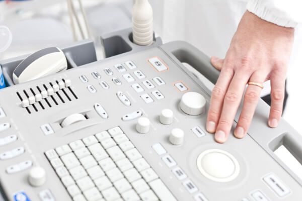
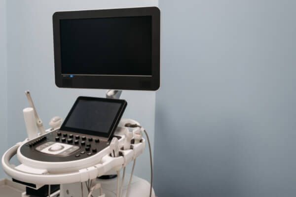
There’s some cost to it like the unit you have, the Sonosite . Nowadays, that’s $8,000, $10,000 or more.
The nice thing is the longer we go down this road, the more competition there is for equipment, the more cost-effective it becomes. There’s some great equipment out there. We use a lot of Sonosite brand ultrasound equipment. There’s Esaote. We’ve got one of their machines that is by far like the Kadlec. It’s so nice. As we’re talking about this, it’s that image quality that improves at each level.
Honestly, if we had a practitioner out there that was interested in talking about how to get on the ground floor and a little bit more of a cost-effective way, there are a few groups out there. I went through HODS, Hands-On Diagnostics , in New York for my training. There are ways within HODS to be more cost-effective, depending on what you’re looking for.
If you’re looking for EMG, there are options out there. There are a few other groups out there that do some of the training. As far as equipment goes, there’s new equipment, then there’s used equipment. There are some good things you can do to make these things available from the ground up to make it a little bit more cost-effective than just swallowing a bunch of debt, which none of us wants to do.
The handheld ones that you can plug into an iPad or your phone or something like that. I think they are called the Butterfly . That’s a couple of thousand dollars? The image quality might not be as good.
I’m an image quality snob now. The more you get into it, the more you’re like, “I want to see that with great quality.” We started with a Lumify, which was one that connected up to an iPad, but you couldn’t do a neural ultrasound if you’re looking at the carpal tunnel and measuring, at least at that time. I’m dating myself because that was a few years ago. Somebody from Lumify is going to be listening to me saying, “We’ve corrected that. We got great caliber for measuring that.” Don’t take that as the gospel truth here. Do your research. There are a lot of options now. Several years ago, when we first started this base, there weren’t a lot of options.
It was Sonosite and that’s it. You either got the $10,000 one, which was the basement model, or you got a $60,000 Kadlec, which was beautiful and took the tests for you. Now, they’re much more affordable. You get what you pay for, essentially. Someone who’s interested in this is going to have to pay for the equipment. It’s going to be a few thousand dollars spent at least. You’re also going to pay for the training.
What I would recommend and I’m sure you would as well, any training would be great as long as there’s additional follow up mentorship after the fact. Having that mentor is almost a must. That could be someone who’s part of the training organization or it could be a physician. I’ve got a friend in New Mexico that’s doing this and he’s got a physician for him that is checking his studies and making sure they’re right.
There are a lot of ways to get to that but you’re right, mentorship is key. Dr. Mohini Rawat is a PT out of New York and works with HODS. He has been my mentor for a number of years. I’ve been doing this for several years and I saw something interesting in a meniscus. I shot her a few snapshots and said, “Mohini, I’m thinking this is this. Am I right?” You like that level of confidence because you need it.
I know it differs state to state and it differs depending upon your Medicare intermediary from what we’re finding out, whether or not Medicare reimburses for it or not. When I started doing an ultrasound in Arizona, my initial claims sent to Blue Cross Blue Shield were denied. I don’t remember what the reason was for denial, but what I had to do was appeal it. I got a letter from the State Board of Physical Therapy saying that ultrasound was within our scope of practice. Based on that appeal with a letter from the Arizona State Board of Physical Therapy, they agreed and then paid for the claims after all.
I had to do the same thing here in Alaska with EMG and ultrasound. Now, I’m getting paid for those here. Depending on the state, you can get reimbursement for an ultrasound but it also depends on the insurance company and your Medicare intermediary. In Arizona, we would get anywhere from $40 up to $200 on ultrasound, outside of your flat ratepayers, which is a separate issue. If my repairs are going to play, you pay a flat rate whatever you do. Does that sound about right? Is that where you’re landing with your range of reimbursement?
Musculoskeletal ultrasound saves patients money and time. It makes it so that you're not doing blind PT.
Click To Tweet
Yeah. The last time that we looked at the pro-ultrasound reimbursement, we were hanging at around $115 across all payers. That’s including somewhere we’re not getting paid anything and somewhere we’re getting paid quite well. That’s been our budget on how we look at things.
That’s where I would hope and wish. Because you’re part of the network with Hands-On Diagnostic, we can work together and push Medicare to recognize physical therapists as providers of musculoskeletal ultrasound and how it benefits the care that we provide. Also, how it lessens the overall cost if we can be more certain about what we’re treating.
We have that discussion with a payer group. The auditor reviewer called us up and said, “I don’t think that a physical therapist should be doing this. This should only be a radiologist.” We said, “How come you think that?” She said, “It’s not like I’m a practitioner. I’m not a provider. It’s because under the classification here, for our insurance company, this comes under a radiology code. I assume since our classification for our insurance company comes under radiology code, only radiologists were doing this.” We said, “Can we tell you how we use it in our practice and show you some research on how this has been used in different states as well as in the military?”
It was so fun because, by the end of the conversation, this individual that initially had a fixed idea that physical therapists should not be doing something with this code said, “You guys have changed my mind.” It’s saving patients’ money. It’s going to save our insurance company money. It’s saving the patient’s time. It’s saving them lost time off work. If we know by screening and doing a musculoskeletal ultrasound on the shoulder, a knee or a hip so that we’re not doing blind PT and waiting for an MRI after we’ve failed at PT. Back to that idea of the order for failed PT.
It’s cheaper than an MRI.
In the end, she flipped and said, “I’m going to take this back. I would like you guys to write a paper for this and give me an essay. Give me all of your bibliography and anything that you can from the military and everything you’ve talked about, the different studies that you’ve brought out, and why this is cost-effective in the hands of the PT. I’m going to go back to the board and I’m going to fight for this because this is in the best patient interest.”
Good luck with that. People who are reading this might want to see where their state lands on this with the State Practice Act. They also have to recognize that as they’re moving forward and doing things that are outside the normal, consider our route to doing dry needling. We have a little bit of resistance to doing dry needling, if you do say so, looking back on our history.
We’re probably going to meet up with some of the same resistance with musculoskeletal ultrasound there. People are going to say, “You can’t play in this sandbox. They’re doing it with us with EMGs.” It should be almost expected at this point as physical therapists to expand what we can do underneath our scope and live within our scope fully.
That’s to be expected, but there are huge benefits to what it can do for our profession. You’re seeing it in your clinics. It’s a great revenue add for you. I know you tied it into what you’re talking about in your book, Debt-Free PT , how you can incentivize your teams and pay back student loans based on what they’re doing with musculoskeletal ultrasound and incentivize them in that way. It’s been a huge boost to your practice. That’s why I love having you on.
I appreciate being able to talk about it. We could keep going for a few hours because this is one of my favorite topics. We’ve hopefully hit on some of the questions and answers from some of the practices, practice owners, and PTs out there that are thinking, “How do I get into this? How do I put my toe in the water and see how this works?”
You’re a pioneer. You’ve said that well, Nathan. There is going to be a little bit of resistance from a handful of people that always want to say, “You can’t do that,” when in reality, Practice Act supports it and everything that you’re doing with mentorship and fellowship and being truly trained and passing that certification exam.
It’s like anything else. It’s like somebody telling you, “You can’t become OCS certified. You shouldn’t do it.” You shouldn’t be on a sideline because there are other practitioners that are there. You’re always going to have your naysayers or whatever, but we’ve always seen physical therapists rise above within our scope and be able to perform. Why? There’s this idea that as a profession, we need to be able to grow and be paid well for what we do because we have a tremendous impact.
We also need to recognize that to get to some of those milestones as a profession. We’re going to be uncomfortable. We’re going to search and we’re going to have to push ourselves to learn at night and study a little bit more, not just put the books away at graduation and never open them up again, but be continually ready to learn and grow.
You used the right word. There’s got to be pioneers that strikeout and are the first to do these things. There are a number of them within the physical therapy industry already. We just need more grassroots, efforts and numbers to get our voice heard.
If we take a look within the APTA today, there are a couple of special interest groups. One within clinical electrodiagnostic wound care. There’s a special interest group in neuromusculoskeletal ultrasound. There’s also another one that is that subset of imaging out of the orthopedics section. There’s a guy in there who might be the vice president and he’s phenomenal.
They have done amazing things and spent their time and their practice in diagnostic ultrasound. We’ve got some leadership that’s going that way. The more physical therapists that open their eyes to what’s the potential in this and what we can do for our practice and for our patients, we’re going to see this be the norm years from now. We’ll see that in all our orthopedic outpatients.
I hope so. If people want to reach out to you and talk to you not only about the musculoskeletal ultrasound but also about your Debt-Free PT book, if they want to look into that, how do they get ahold of you?
A couple of ways. My email address is BartMPT@Gmail.com. You can always shoot me an email and ask questions. I usually have about a three-day lag. I try to keep up with my email. Usually, within about 3 to 5 days, I’ll get back to you and we’ll get something set up. Even my cell phone number, I don’t answer it during the day but if you want to shoot me a call or a text, I’ll throw that out there. We might get bombarded this way. I’ll try to get back to that one.
My cell phone number is (208) 705-4678. Leave me a message and we’ll try to get you to the right place. I’m so excited about where our profession is going, especially at a time when we have a lot of doomsayers and naysayers out there that decrease reimbursement. I don’t know where the future of PT is, but the future of PT is now. It’s here and a big part of it is in diagnostics.
If people want to read your book, do you have a landing page?
Yes. It’s www.Debt-FreePT.com.
Good book. I love it. Thanks for your time, Bart. I appreciate it.
Thanks, Nathan. We’ll talk to you later.
Take care.
Important Links
About Bart McDonald
 Bart McDonald, PT, MPT, ECS, FMSK, is the President and owner of Superior Physical Therapy. Bart began his career at an outpatient clinic in Montana. He later worked at Bannock Regional Medical Center.
Bart started Aspen Physical Therapy and later decided to open his own clinic, Superior Physical Therapy in 2008 which has grown to four clinics in southeast Idaho.
Bart graduated with his Master’s of Physical Therapy from Emory University School of Medicine in 2000. He specializes in knee, shoulder, and spine rehabilitation, Electromyography and Nerve Conduction Study testing and is ASTYM certified. He is Board Certified in Clinical Electrophysiology and is a Fellow in Musculoskeletal Ultrasound.
Bart grew up in Nampa, Idaho, is married and has three children and one grandchild. When he’s not working, he is spending time with his family, water skiing, or downhill skiing.